Search
Dr Jessica Buck and Associate Professor Raelene Endersby have been appointed to the prestigious Australian Brain Cancer Mission Expert Advisory Panel.
Associate Professor Lesterhuis said the gel, developed with the help of chemists at The University of Western Australia, could revolutionise the way solid tumours were treated.
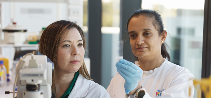
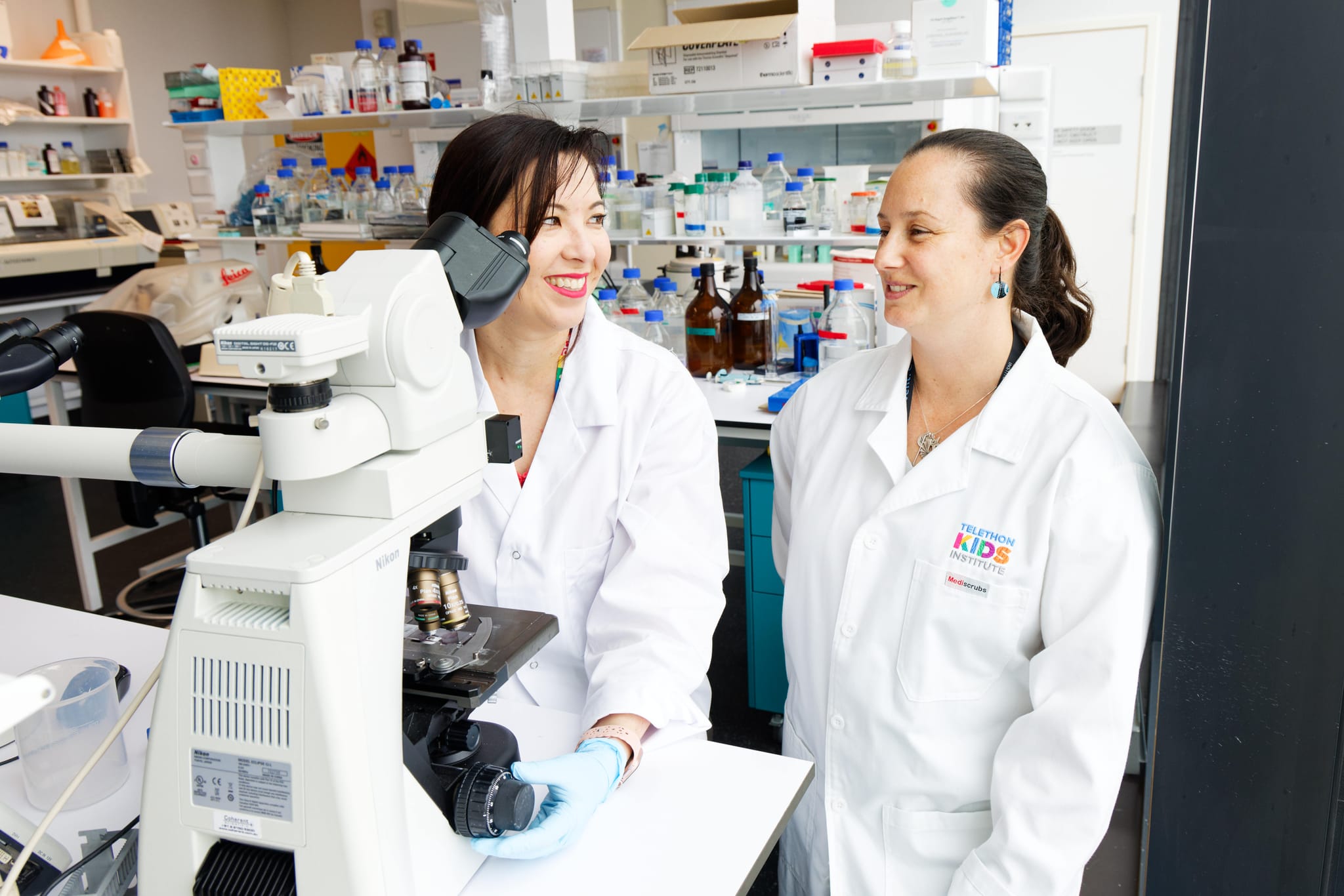
The WA Kids Cancer Centre has a suite of world-leading research projects to unlock new treatments for childhood cancers.
High-grade gliomas including glioblastoma (GBM) and diffuse midline gliomas (DMG) represent the most lethal and aggressive brain cancers where current treatment modalities offer limited efficacy. Chimeric antigen receptor (CAR) T cell therapies have emerged as a promising strategy, boasting tumor-specific targeting and the unique ability to penetrate the blood-brain barrier.
Early, rapid, and accurate diagnostic tests play critical roles not only in the identification/management of individuals infected by SARS-CoV-2, but also in fast and effective public health surveillance, containment, and response. Our aim has been to develop a fast and robust fluorescence in situ hybridization (FISH) detection method for detecting SARS-CoV-2 RNAs by using an HEK 293 T cell culture model.
An in-depth investigation of gene regulation and cell populations at sites of fetal blood-cell production provides clues as to why children with Down’s syndrome are predisposed to developing leukaemia.
T cells engineered to express chimeric-antigen receptors (CAR-T cells) can effectively control relapsed and refractory haematological malignancies in the clinic. However, the successes of CAR-T cell therapy have not been recapitulated in solid tumours due to a range of barriers such as immunosuppression, poor infiltration, and tumour heterogeneity.
There are few options for patients with relapse/refractory B-cell acute lymphoblastic leukemia, thus this is a major area of unmet medical need. Here, we reveal that inclusion of a poison exon in RBM39, which could be induced both by CDK9 or CDK9 independent CMGC (cyclin-dependent kinases, mitogen-activated protein kinases, glycogen synthase kinases, CDC-like kinases) kinase inhibition, is recognized by the nonsense-mediated mRNA decay pathway for degradation.
Despite significant advances, outcomes for children with Down syndrome (DS, trisomy 21) who develop acute lymphoblastic leukemia remain poor. Reports of large DS-ALL cohorts have shown that children with DS have inferior event-free survival and overall survival compared to children without DS.
Gliomas account for nearly 30% of all primary central nervous system (CNS) tumors in children and adolescents and young adults (AYA), contributing to significant morbidity and mortality. The updated molecular classification of gliomas defines molecularly diverse subtypes with a spectrum of tumors associated with age-distinct incidence.